枕叶及其功能

本文转载内容网址:https://human-memory.net/occipital-brain-lobe/
本文仅作翻译文章,翻译内容仅供参考,本文遵守CC-BY-SA许可协议将译文发布在个人博客上(7988888.xyz),仅供学术参考使用,英文原文知识版权归原作者所有,此文章中所有链接均通过博客进行访问。
The occipital lobe participates in vision processing. It processes and interprets everything we see. The occipital lobe is also responsible for analyzing contents, such as shapes, colors, and movement, and also for interpreting and drawing conclusions about the images we see.
枕叶参与视觉处理。它处理和解释我们看到的一切。枕叶还负责分析内容,例如形状、颜色和运动,还负责解释和总结关于我们看到的图像。
Boundaries, Anatomy, Position, and Structure of the Occipital Brain Lobe枕叶的边界、解剖、位置和结构
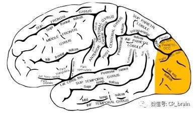
The boundaries of the occipital lobe include the edges of the parietal and temporal lobe. The occipital lobe contains the primary visual cortex and associative visual areas (1).
枕叶的边界包括顶叶和颞叶的边缘。枕叶包含初级视觉皮层和相关视觉区域 ( 1 )。
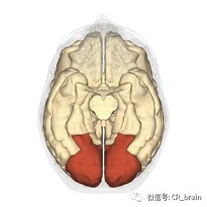
The occipital lobe occupies the posterior parts of the hemispheres. On the convex surface of the hemisphere, the occipital lobe has no sharp boundaries separating it from the parietal and temporal lobes.
枕叶位于大脑半球的后部。在大脑半球的凸面上,枕叶与顶叶和颞叶之间没有明显的界限。
The exception is the upper part of the parietal-occipital groove, which, located on the inner surface of the hemisphere, separates the parietal lobe from the occipital lobe. The furrows and edges of the upper canopy of the occipital lobe are unstable and have a variable structure.
例外的是顶枕沟的上部,它位于大脑半球的内表面,将顶叶和枕叶分开。枕叶上冠的沟和边缘不稳定,结构多变。
On the inner surface of the occipital lobe, there is a groove of spores, which separates the wedge (triangular norm of the occipital lobe) from the lingual gyrus and the occipital-temporal gyrus (1).
在枕叶的内表面,有一个孢子沟,将楔形(枕叶三角线)与舌回和枕颞回分开(1)。
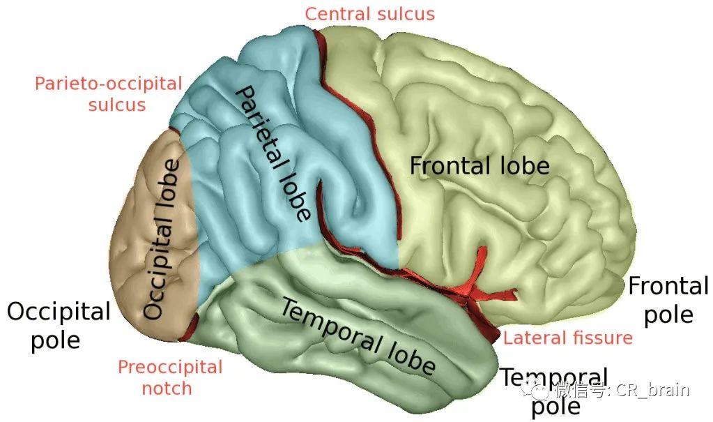
In the occipital lobe of the cerebral cortex is, the following fields are positioned:
在大脑皮层的枕叶中,定位了以下位置:
Area 17 - Gray matter buildup in a visual analyzer. This field is the primary zone. It is made up of 300 million nerve cells.视觉分析中的灰质堆积。该位置是主要区域。它由3亿个神经细胞组成。
Area 18 - It is also a nuclear set of visual analyzers. This field performs the function of perceived writing and is a more complex secondary area. 它也是一组核心的视觉分析。这个位置执行感知写作的功能,是一个更复杂的次要领域
Area 19 – This field is involved in evaluating the value of what we see. 该位置涉及评估我们所看到的事物的价值。
Area 39 - This brain part does not completely belong to the occipital region. This area is located at the border between the parietal, temporal, and occipital lobes. Its functions include integrating visual, auditory, and general sensitivity of information (1).这个大脑部分并不完全属于枕区。该区域位于顶叶、颞叶和枕叶之间的边界处。它的功能包括整合信息的视觉、听觉和一般敏感性 ( 1 )。
The Function of the Occipital Brain Lobe枕叶功能
The function of the occipital lobe is related to the perception and processing of visual information, as well as the organization of complex processes of visual perception.枕叶的功能与视觉信息的感知和加工处理,以及复杂的视觉感知过程的组织有关。
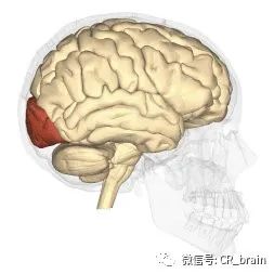
At the same time, the upper half of the retina, which detects light from the lower field of vision, is projected into a wedge; in the reed area, the gyrus is the lower half of the retina, noticing the light from the upper field of view(1).
同时,视网膜的上半部分,检测来自下视野的光,被投射到楔形物中;在 reed 区,脑回是视网膜的下半部分,注意到来自上视野的光线(1).
In the occipital cortex, there is a primary visual area (the cortex of the part of the sphenoid gyrus and the lingual lobule). There is a local representation of the retinal receptors. Each retinal point corresponds to its portion of the visual cortex, while the yellow dot area has a relatively large representation area.
在枕叶皮层,有一个初级视觉区(蝶骨回和舌小叶部分的皮层)。有视网膜受体的局部表现。每个视网膜点对应其视觉皮层的一部分,而黄点区域则具有相对较大的表示区域。
In connection with the incomplete intersection of visual pathways, the same half of the retina is projected into the visual area of each hemisphere. The presence of retinal projection of both eyes in each hemisphere is the basis of binocular vision.
由于视觉通路的不完全交叉,同一半的视网膜投射到每个半球的视觉区域。双眼在每个半球的视网膜投影是双眼视觉的基础。
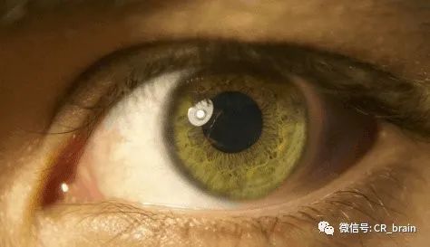
The neurons of these zones are polymodal and respond not only to light but also to tactile and auditory stimuli. Different types of sensitivities are synthesized in this visual area, more complex visual images emerge and they are recognized.
这些区域的神经元是多模态的,不仅对光有反应,而且对触觉和听觉刺激也有反应。在这个视觉区域中合成了不同类型的敏感性,产生了更复杂的视觉图像,并对其进行识别。
An example can be given to understand the function of the occipital lobe in visual perception. When we look at a map and “put” the route planner information into our working memory, the first step would be to process millions of light stimuli in the recognition centers, i.e. to process different signals perceived by the photosensitive cells of our retina.
通过实例说明枕叶在视觉知觉中的作用。当我们查看地图并将路线规划信息“放入”到工作记忆中时,第一步是处理识别中心中数百万个光刺激,即处理视网膜光敏细胞感知的不同信号。
Furthermore, the occipital lobe receives incoming information, which is processed and immediately sent to the hippocampus, where it is formed into memory. Firstly, it becomes short-term memory. Accordingly, we remember the name of the place of destination and remember it as we move along this route.
此外,枕叶接收传入的信息,这些信息被处理并立即发送到海马体,在那里形成记忆。首先,它变成了短期记忆。因此,我们记住目的地的名称,并在沿着这条路线移动时记住它。
As we have already stated, the occipital lobe is responsible for the visual perception of information, as well as its operational storage. Generally, everything projected by the retina is recognized and formed into a specific image in the occipital lobe.
正如我们已经说过的,枕叶负责信息的视觉感知及其操作存储。一般来说,视网膜投射的一切都会在枕叶中被识别并形成特定的图像。
For absolutely healthy people, this proportion works independently and flawlessly, but irreparable consequences can occur with injuries and some illnesses. Sometimes, total blindness can occur. This is the process that happens if there is a damage on the surface of the primary visual cortex.
对于绝对健康的人来说,这个比例可以独立且完美地发挥作用,但受伤和某些疾病可能会导致无法弥补的后果。有时,可能会发生完全失明。如果初级视觉皮层表面受损,就会发生这个过程。
Light signals transmit information to the occipital lobe via nerve endings, which represents a form of irritation or stimuli for the retina. The nerves then transmit information to the diencephalon, another part of the brain. And diencephalon, in turn, sends information to the primary visual cortex, called the sensory cortex.
光信号通过神经末梢将信息传输到枕叶,这代表了对视网膜的一种刺激或刺激形式。然后神经将信息传输到大脑的另一部分间脑。间脑将信息发送到初级视觉皮层,称为感觉皮层。
From the primary sensory cortex, nerve signals are sent to the adjacent areas and are called sensory associative cortex areas. The main function of the occipital lobe is to send signals from the primary visual cortex to the visual associative cortex. The areas described together analyze the visual information observed and retain visual memories.
从初级感觉皮层向相邻区域发送神经信号,称为感觉联想皮层区。枕叶的主要功能是将信号从初级视觉皮层发送到视觉联想皮层。所描述的区域一起分析观察到的视觉信息并保留视觉记忆。
As already implied, this occurs when the primary visual cortex, whose surface is visible, is damaged. Complete damage to the primary cortex occurs in three cases, as a result of a head injury, as a result of the development of a tumor on the surface of the brain, and finally, yet very rarely, as a consequence of certain congenital anomalies.
当表面可见的初级视觉皮层受损时,就会发生这种情况。在三种情况下会发生初级皮质的完全损伤,头部受伤、大脑表面肿瘤的发展、某些先天性异常。
Damage of the Occipital Brain Lobe枕叶损伤
Damage to the primary visual cortex leads to a form of central blindness called the Anton’s syndrome; patients cannot recognize objects via their sense of sight and are completely unaware of their deficits (2).
初级视觉皮层的损伤会导致一种称为安东综合征的中枢性失明;患者无法通过视觉识别物体,并且完全没有意识到自己的缺陷 ( 2 )。
Epileptic seizures in the area of the occipital lobe cause visual hallucinations, most commonly in the form of dashes and a colored mesh that appears on the contralateral field of view.
枕叶区域的癫痫发作会导致视觉幻觉,最常见的形式是出现在对侧视野上的破折号和彩色网格。
Damage of the occipital brain lobe can occur as a result of a head injury, a tumor on the surface of the brain, and certain congenital anomalies.
头部受伤、大脑表面的肿瘤和某些先天性异常也可能会导致枕叶脑区受损。
However, focal lesions do not lead to complete loss of vision. For example, after taking a familiar object in his hands, a patient may describe the object he/she is touching. However, if that same object is shown in the picture, then the patient will not be able to describe its shape and color. In medical language, this condition is called visual agnosia.
局灶性病变不会导致视力完全丧失。例如,患者将熟悉的物体拿在手中后,可能会描述他/她正在触摸的物体。但是,如果图片中显示相同的物体,则患者将无法描述其形状和颜色。在医学语言中,这种情况称为视觉失认症。
At times, focal lesions can localize and restore vision and perception. However, it is important to note that the chances of a partial recovery in children are higher than those in patients whose brain is already formed and not growing anymore. Treatment is usually done surgically(2).
有时,局灶性病变可以定位并恢复视力和知觉。然而,重要的是要注意,儿童部分康复的几率高于大脑已经形成且不再生长的患者。治疗通常通过手术进行(2)。
Pain in the occipital brain lobe region枕叶区域疼痛
There are many different causes of pain in this region. Some of them include:
该区域有许多不同的疼痛原因。其中一些包括:
Nerve tension and stress. With prolonged tension, neck and back muscle spasms and neck pain occurs. Also, pain in the occipital brain region can be localized. The patient can diminish the pain by breathing calmly and deeply. If the pain does not stop after the patient feels relaxed, a visit to a doctor is obligatory. 随着长期紧张,颈部和背部肌肉痉挛和颈部疼痛发生。此外,枕叶大脑区域的疼痛可以被定位。患者可以通过平静深呼吸来减轻疼痛。如果患者感到放松后疼痛仍未停止,则必须去看医生。
Osteochondrosis of the cervical spine. This condition results in sharp pain in the back of the head. Specialized forms of gymnastics can help. However, the patient must see a neurologist. 这种情况会导致后脑勺剧烈疼痛。特殊形式的体操可以提供帮助。但是,患者必须去看神经科医生。
High blood pressure. This condition can cause pain with a feeling of fullness. Pressure control is essential for extending one’s lifetime. Contact a neurologist if you feel pain in the occipital part of your brain and suffer from blood pressure disorders. 这种情况会导致饱腹感疼痛。压力控制对于延长寿命至关重要。如果您感到大脑枕部疼痛并患有血压紊乱,需要神经科医生查看。
Increased intracranial pressure. This serious condition is characterized by oppressive eye pain. The pain is localized in the occipital lobe. The patient must immediately see a doctor.这种严重疾病的特点是压迫性眼痛。疼痛位于枕叶。患者必须去看医生。
Conclusion结论
The occipital lobe is located in a triangle, the apex of which is the parietal lobe and the sides of the temporal lobes of the brain. The cerebellum is positioned below the occipital lobe. This brain part has a variable structure.
枕叶位于一个三角形中,其顶点是顶叶和大脑颞叶的两侧。小脑位于枕叶下方。这个大脑部分具有可变结构。
Its key function is processing visual information. The visual cortex, located on both hemispheres of the occipital lobe, provides binocular vision - the world seems vast and wide to the human eye.
它的主要功能是处理视觉信息。视觉皮层位于枕叶的两个半球,提供双目视觉——在人眼看来,世界是广阔的。
The visual cortex, called the associative area, constantly communicates with other brain structures, forming a complete image of the world. The occipital lobe has strong links with the limbic system (especially the hippocampus), the parietal, and temporal lobes.
视觉皮层,称为联想区,它不断地与其它大脑结构进行交流,形成一个完整的世界图像。枕叶与边缘系统(尤其是海马体)、顶叶和颞叶有很强的联系。
Thus, this or that visual image may be accompanied by negative emotions or vice versa: long-term visual memory may evoke positive emotions.
因此,这个视觉图像可能伴随着消极情绪,反之亦然:长期的视觉记忆可能会唤起积极的情绪。
The occipital lobe, along with simultaneous signal analysis, also plays the role of an information container. However, the amount of such information is insignificant, and most of the environmental information is stored in the hippocampus.
枕叶与同步信号分析一起,也扮演着信息容器的角色。但是,这类信息量微乎其微,大部分环境信息都存储在海马体中。
The occipital cortex is strongly associated with feature integration. The essence of these theories is that the cortical analytic centers of separate properties of an object (color) are processed separately, and in parallel with the processing of other information.
枕叶皮质与特征整合密切相关。这些理论的本质是,一个对象(颜色)的独立属性的大脑皮层分析中心是分开处理的,并与其它信息的处理并行。
To sum up, the occipital lobe is responsible for processing visual information and their integration into the general relation to the world; storing visual information; interaction with other areas of the brain, and, partly, tracking their functions; as well as the binocular perception of the environment.
综上所述,枕叶负责处理视觉信息并将其整合到与世界的一般关系中;存储视觉信息;与大脑的其它区域相互作用,并在一定程度上跟踪它们的功能;以及对环境的双目感知。
References参考:
Rehman A, Al Khalili Y. Neuroanatomy, Occipital Lobe. [Updated 2019 Jul 6]. In: StatPearls [Internet]. Treasure Island (FL): StatPearls Publishing; 2019 Jan-. Available from: https://www.ncbi.nlm.nih.gov/books/NBK544320/ Found online at: https://www.ncbi.nlm.nih.gov/books/NBK544320/
Macaskill J. A CASE OF OCCIPITAL LOBE INJURY. Br J Ophthalmol. 1945 Dec;29(12):626-8. PMID: 18170164; PMCID: PMC512175. Found online at: *https://www.ncbi.nlm.nih.gov/pmc/articles/PMC512175/
谢谢大家观看,如有帮助,来个喜欢或者关注吧!
本文作者:human-memory
译文作者:陈锐
博客地址 :陈锐博客
知乎地址 : 知乎专栏
B站地址 : B站主页
书店地址 : 书店主页
CSDN地址 : csdn主页
仅供学习使用,不作其它用途,如有侵权,请留言联系,作删除处理!
